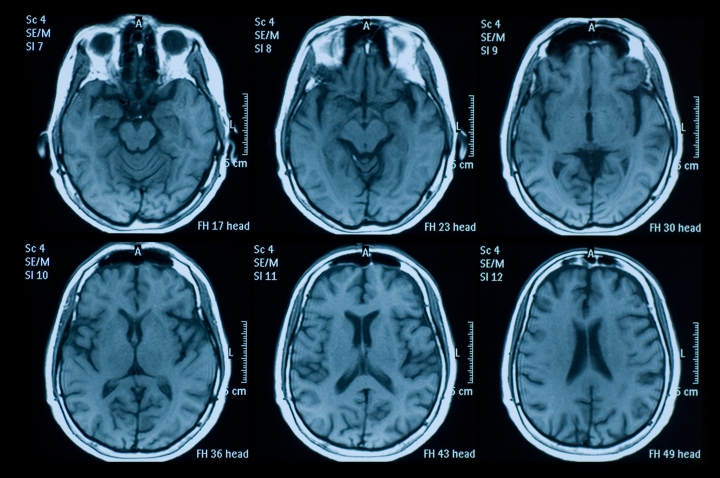
Since the development of functional magnetic resonance imaging in the 1990s, the reliance on neuroimaging has skyrocketed as researchers investigate how fMRI data from the brain at rest, and anatomical brain structure itself, can be used to predict individual traits such as depression, cognitive decline, and brain disorders.
Brain imaging has the potential to reveal the neural underpinnings of many traits, from disorders like depression and chronic widespread pain to why one person has a better memory than another, and why some people’s memories are resilient as they age. But how reliable brain imaging is for detecting traits has been a subject of wide debate.
Prior research on brain-wide association studies, known as BWAS, has shown that links between brain function and structure and traits are so weak that thousands of participants are needed to detect replicable effects. Research of this scale requires millions of dollars in investment in each study, limiting which traits and brain disorders can be studied.
However, according to a new commentary published in Nature, stronger links between brain measures and traits can be obtained when state-of-the-art pattern recognition (or ‘machine learning’) algorithms are utilized, which can garner high-powered results from moderate sample sizes.
In their article, researchers from Dartmouth and University Medicine Essen provide a response to an earlier analysis of brain-wide association studies led by Scott Marek at Washington University School of Medicine in St. Louis, Brenden Tervo-Clemmens at Massachusetts General Hospital/Harvard Medical School, and colleagues. The earlier study found very weak associations across a range of traits in several large brain imaging studies, concluding that thousands of participants would be needed to detect these associations.
The new article explains that the very weak effects found in the earlier paper do not apply to all brain images and all traits, but rather are limited to specific cases. It outlines how fMRI data from hundreds of participants, as opposed to thousands, can be better leveraged to yield important diagnostic information about individuals.
One key to stronger associations between brain images and traits such as memory and intelligence is the use of state-of-the-art pattern recognition algorithms. “Given that there’s virtually no mental function performed entirely by one area of the brain, we recommend using pattern recognition to develop models of how multiple brain areas contribute to predicting traits, rather than testing brain areas individually,” says senior author Tor Wager, the Diana L. Taylor Distinguished Professor of Psychological and Brain Sciences and director of the Brain Imaging Center at Dartmouth.
“If models of multiple brain areas working together rather than in isolation are applied, this provides for a much more powerful approach in neuroimaging studies, yielding predictive effects that are four times larger than when testing brain areas in isolation,” says lead author Tamas Spisak, head of the Predictive Neuroimaging Lab at the Institute of Diagnostic and Interventional Radiology and Neuroradiology at University Medicine Essen.
However, not all pattern recognition algorithms are equal, and finding the algorithms that work best for specific types of brain imaging data is an active area of research. The earlier paper by Marek, Tervo-Clemmens, and others also tested whether pattern recognition can be used to predict traits from brain images, but Spisak and colleagues found that the algorithm they used is suboptimal.
When the researchers applied a more powerful algorithm, the effects got even larger and reliable associations could be detected in much smaller samples. “When you do the power calculations on how many participants are needed to detect replicable effects, the number drops to below 500 people,” Spisak says.
“This opens the field to studies of many traits and clinical conditions for which obtaining thousands of patients is not possible, including rare brain disorders,” says co-author Ulrike Bingel at University Medicine Essen, who is the head of the University Centre for Pain Medicine. “Identifying markers, including those involving the central nervous system, are urgently needed, as they are critical to improve diagnostics and individually tailored treatment approaches. We need to move towards a personalized medicine approach grounded in neuroscience. The potential for multivariate BWAS to move us towards this goal should not be underestimated.”
The team explains that the weak associations found in the earlier analysis, particularly through brain images, were collected while people were simply resting in the scanner, rather than performing tasks. But fMRI can also capture brain activity linked to specific moment-by-moment thoughts and experiences.
Wager believes that linking brain patterns to these experiences may be a key to understanding and predicting differences among individuals. “One of the challenges associated with using brain imaging to predict traits is that many traits aren’t stable or reliable. If we use brain imaging to focus on studying mental states and experiences, such as pain, empathy, and drug craving, the effects can be much larger and more reliable,” says Wager. “The key is finding the right task to capture the state.”
“For example, showing images of drugs to people with substance use disorders can elicit drug cravings, according to an earlier study revealing a neuromarker for cravings,” says Wager.
“Identifying which approaches to understanding the brain and mind are most likely to succeed is important, as this affects how stakeholders view and ultimately fund translational research in neuroimaging,” says Bingel. “Finding the limitations and working together to overcome them is key to developing new ways of diagnosing and caring for patients with brain and mental health disorders.”