Microscopes are indispensable in disease diagnosis and are employed every day by pathologists who magnify slivers of tissue to detect telltale signs of infection, blood disorders, and cancers.
Now, instead of using samples mounted on glass slides, a new smart microscope created by Dartmouth Health dermatopathologist Aravindhan Sriharan and students at the Digital Applied Learning and Innovation Lab will allow clinicians to diagnose disorders from digital images.
By combining the brains of a computer with the design of a medical microscope, RavaOne: The SmartScope is poised to integrate traditional microscopy with rapidly emerging health care technologies that rely on artificial intelligence and expand the possibilities of telemedicine, says Sriharan, who is an assistant professor of pathology and laboratory medicine at the Geisel School of Medicine.
Like so many other fields, pathology is on the verge of being transformed by AI algorithms that can make diagnoses more accurate, streamlined, and affordable. To make the leap towards adopting these tools, pathologists must make the switch to working off digital images.
A trained pathologist who would take just a few seconds to make a routine diagnosis using a traditional microscope would likely spend several minutes scrolling through high-resolution images on a monitor if they were using a computer alone.
“Doing digital pathology on a computer screen is incredibly onerous,” says Sriharan. “Medical microscopes have been iterated over 200 years to do one thing really well, but computer monitors are generalists,” he says.
To Sriharan, the solution lay in combining a computer with a microscope in action, akin to how a smartwatch merges functions.

“A smartwatch has the form factor of a wristwatch on the outside but is essentially a computer on the inside. And it can do things that neither a computer nor a wristwatch acting alone can do,” says Sriharan, who pitched his idea to the DALI Lab.
Lauren Goyette ’23, an engineering major with an interest in product design, began working on the project as a designer last fall.
“Pathologists are so efficient with microscopes; it’s very impressive,” she says. The challenge, as Goyette saw it, was to help them work at speed as they made the transition to the digital microscopy world. She worked on creating the SmartScope’s chassis using 3D printing.
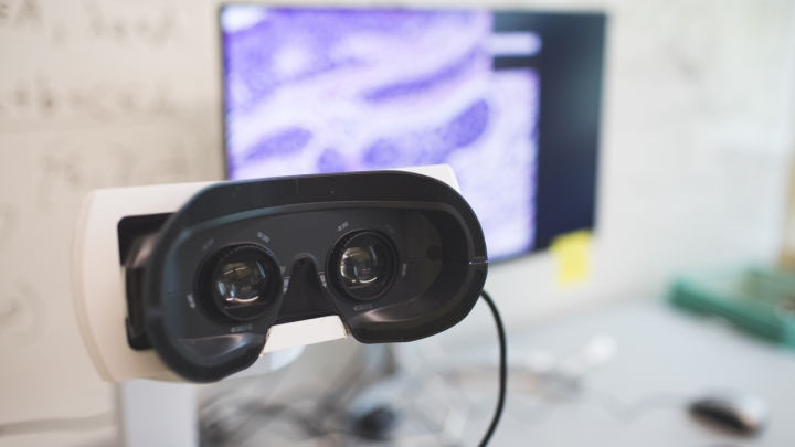
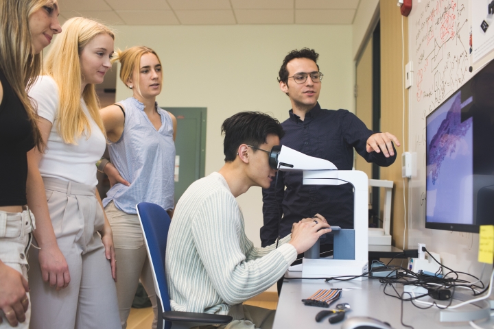
Complete with an eyepiece and a stage for the glass slide to sit on, the SmartScope preserves the familiar form of a laboratory microscope.
What’s different is that in place of a laboratory slide with stained tissue, the SmartScope stage holds a “dummy” slide. As a user moves this slide, a camera and computer system track its position. This movement is linked to the digitized, high resolution picture of a tissue sample, which moves as the user looks through the eyepiece, mirroring the experience of a conventional microscope.
“Just getting an image to display was a major milestone,” says Alex Carney ’23, who has also been with the project all along as a developer. Subsequently, he worked on getting the large image file to move smoothly across the user’s field of vision as they moved the glass slide on the stage.
“The goal is to make a product that someone would be comfortable using for multiple hours a day to do real work—real research and diagnostics. We’re still working on that and we’re very close, but that was the biggest challenge of this project,” says Carney, who didn’t anticipate how much the project would sharpen his math and software skills when he began.
The team received positive reviews from users who tested their prototype in the winter term.
“There was a lot of thought and detail put into the simplistic, yet highly advanced intentions of the SmartScope, while maintaining the essential functionalities of a microscope,” says Jessica Bentz, assistant professor of pathology affiliated with Dartmouth Hitchcock and the Geisel School of Medicine.
“I was impressed by the clarity of images at each magnification,” says Bentz, who called attention to the SmartScope’s turnstile that allows users to switch between magnifications—a more convenient replacement for the rotating carousel of lenses that traditional microscopes use for this purpose. It was clearly designed with an ergonomic advantage for the user, she says.
“The SmartScope skillfully marries the growing impact of digital imaging and AI as a tool for diagnostic pathology with the familiarity of sitting at a microscopic instrument,” says Bentz.
“This technology has the potential to change diagnostics by allowing providers across the world to collaborate instantaneously,” says Jorie MacDonald ’25, a computer science major who joined the team as a project manager in spring.
“You wouldn’t need to ship a physical slide, hoping that it gets through customs and reaches the lab without getting damaged somehow; you just send a file,” she says.

The device is patent pending, and will form the cornerstone of PixCell, a new startup incorporated by Sriharan. “The SmartScope can finally actualize the promise of AI in disease diagnostics,” he says.
Sriharan believes that the technology can transform health care not just in countries that are leading the change but also in resource- and expertise-limited regions. The device would help providers on the ground screen cases to identify the ones that need immediate attention, sending them on to colleagues and volunteers across the globe.
Pathologists can run AI algorithms to simulate the results of expensive and time-consuming laboratory tests on the images they examine, saving patients time and money. The algorithms can also help chart the likely course of the disease and recommend effective treatments.
“We can actually bring first-world medicine—not watered down, but high-tech and cutting edge—to people who need it most,” says Sriharan.
Other members of the DALI team who worked on developing the RavaOne: SmartScope include Andy Kotz ’24, Ziray Hao ’22, Atharv Agashe ’25, Elizabeth Frey ’24, Victor Muturi ’23, Lauren Kidman ’25, Daniel Lubliner ’25, Annie Qiu ’24, Emily Chen ’24, Ulgen Yildirim ’24, Alejandro Lopez ’23, Joy Miao ’23, and Kelly Song ’23.

