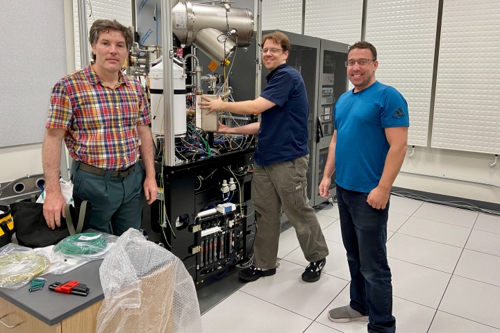
A versatile electron microscope is being installed in a specially built laboratory in the Class of 1982 Engineering and Computer Science Center and will be ready for use this fall.
The Thermo Scientific Talos F200i scanning transmission electron microscope will afford researchers, both at Dartmouth and other universities in northern New England, a clearer view of materials at the atomic level.
“The new field emission gun scanning transmission electron microscope replaces an aging 20-year-old TEM. It provides us higher resolution imaging, higher resolution X-ray spectroscopy, and new imaging capabilities,” says Ian Baker, the Sherman Fairchild Professor of Engineering at Thayer School of Engineering, who was awarded a $1 million National Science Foundation grant that helped Dartmouth purchase the instrument.
Baker studies defects in materials that affect and shape their mechanical, electrical, or magnetic properties. Understanding microstructure helps improve material properties, Baker says. “If you don’t know what it looks like, you will simply be playing in the dark.”
Transmission electron microscopes illuminate objects with electrons to reveal their structures at incredibly high resolution, capturing images of individual molecules or even atoms that make up the specimen under study.
Scanning TEMs expand the capabilities of conventional TEMs by using a tightly focused, nanometer-sized beam of electrons to scan the sample, one small area at a time. This produces X-rays that are specific to the elements present in the sample area, allowing researchers to map the material’s chemical composition.
“This is the first Talos F200i in the U.S. to be equipped with a cold field emission gun,” says Electron Microscope Facility Director Maxime Guinel de France. This emitter, which shoots out electrons at the specimen, allows faster imaging with better resolution and can be adjusted to lower voltage levels when sensitive materials are being studied.
“It’s a really exciting development for Dartmouth,” says Dean Madden, vice provost for research and professor of biochemistry and cell biology.
Madden believes that the instrument will find users from across campus—material scientists in engineering, physics, and chemistry as well as biologists and medical researchers who can use it to study the structures of proteins. He also notes that more than 75% of the hands-on research on the current instrument is performed by students, a trend that is likely to continue with the Talos.
For some types of advanced imaging, like cryogenic electron microscopy, scientists carry their samples to national laboratories that house the necessary equipment. In these cases, “our hope is that the Talos microscope will serve as a springboard where researchers can do preliminary experiments and get ready to then go out to national facilities for the really high-resolution pictures,” Madden says.
The new instrument also comes with remote monitoring and operation capabilities that will make it accessible to off-site users for courses and research projects. The NSF proposal outlined plans to extend the use of the scanning TEM to researchers at University of Maine-Orono, University of Vermont, and University of New Hampshire, as well as a statewide network of colleges and universities.
“The Talos microscope is the only instrument of its kind in this region,” says Baker. “It will make possible numerous research projects requiring high-resolution imaging and elemental analysis.”

